fig 45. Transvaginal image of the uterus of an 85-year-old woman reveals calcification in the arcuate arteries (arrows)..gif
-
Download this image
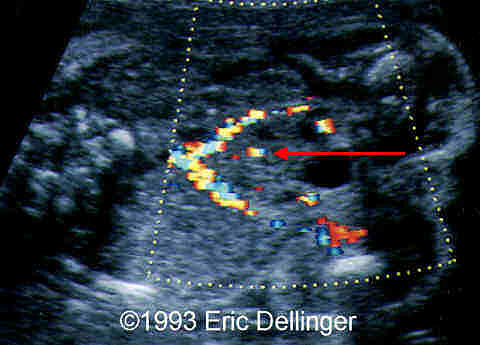
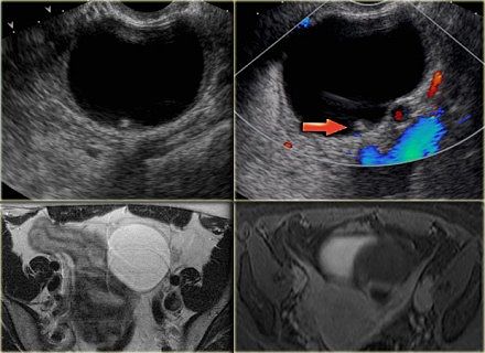
FIG 72 The pelvic kidney is seen between the iliac vessels in the upper portion of the image. The color Doppler demonstrates a renal artery (red arrow)..jpg
-
Download this image
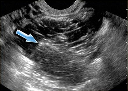
FIG 71.jpg
-
Download this image
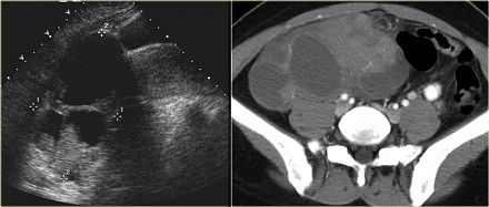
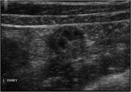
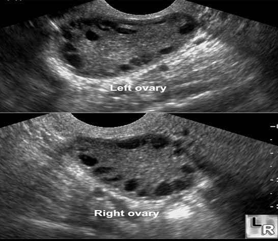
FIG 68On ultrasound both ovaries are markedly enlarged and contain cystic components with intracystic solid components.jpg
-
Download this image
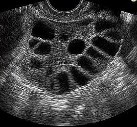
FIG 66.jpg
-
Download this image
FIG 70Mature cystic teratoma with a Rokitansky nodule or dermoid plug.jpg
-
Download this image
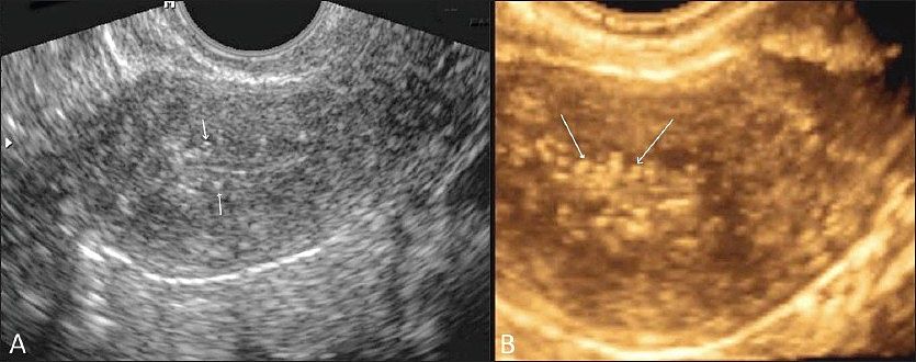
FIG 64 PIDPelvic inflammatory disease. Transvaginal 2D gray-scale (A) and 3D color (B) USG images show subendometrial calcification (white arrows).jpg
-
Download this image
FIG 62.gif
-
Download this image
FIG 60.jpg
-
Download this image
FIG 59.jpg
-
Download this image
Next